Breastmilk carries more than nutrition


New research explores how tiny particles in breastmilk may carry molecular signals from mother to infant — and what this could mean for early brain development.
Breastmilk does more than provide nutrition. It contains a range of biological components that may help support an infant’s developing immune system and other organs. Researchers are now exploring whether it also carries molecular messages that influence how the developing brain works.
A 2026 study published in Cell Metabolism suggests that postnatal depression (PND) may be associated with changes in some of these messages. The researchers found that breastmilk from mothers with PND contained different levels of particular microRNAs (miRNAs), small molecules that help regulate gene activity. In experiments involving mice, some of these miRNAs were linked to changes in brain-cell development and learning and memory.
The findings offer a possible biological explanation for how a mother’s postnatal mental health may influence an infant’s development. However, it is important to distinguish what the study demonstrated in mice from what is currently known about human babies.
What are extracellular vesicles, and why are they important?
Cells release tiny, membrane-covered particles called small extracellular vesicles (sEVs). These particles can carry biological material, including miRNAs, from one cell to another. Their membrane helps protect this cargo and may allow it to be delivered to other cells.
MiRNAs do not usually make proteins themselves. Instead, they help regulate gene expression by interacting with messenger RNA, which carries instructions used by cells to make proteins. In this way, miRNAs can influence which proteins a cell produces and how it functions.
The researchers asked whether sEVs in breastmilk could carry miRNAs to an infant’s brain, and whether changes in these miRNAs might help explain some of the effects associated with maternal PND.
To investigate this relationship, the researchers employed a mouse model of chronic unpredictable mild stress (CUMS) to simulate maternal stress. They used offspring born to stressed dams (CUMS dams) and comparing them with offspring from healthy control dams (HC dams). Both male and female offspring were included in the experimental design. Cross-fostering was performed between the two groups, whereby offspring born to CUMS dams were raised by healthy control dams and vice versa. This experimental approach enabled the researchers to distinguish the potential effects of the postnatal maternal environment, particularly breastfeeding and maternal care, from prenatal influences associated with maternal stress. Offspring raised by their biological dam-type counterparts (CUMS pups with CUMS dams and HC pups with HC dams) served as control groups. The effects of maternal stress on offspring cognitive and emotional development were subsequently evaluated using a series of established behavioural assays, as summarised in the table below:
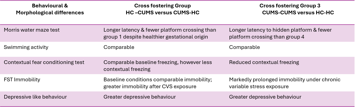
The findings revealed that offspring born to healthy mothers but subsequently fostered by mothers exhibiting postpartum depression (PND)-like symptoms developed significant cognitive and emotional impairments. In contrast, offspring born to stressed mothers but fostered by healthy mothers showed substantially fewer behavioural abnormalities. These findings suggest that the postnatal maternal environment plays a critical role in offspring neurodevelopment, independent of prenatal exposure to maternal stress. Notably, when sex differences were considered, these behavioural deficits were predominantly observed in male offspring.
To investigate the neurobiological mechanisms underlying these observations, the researchers conducted a series of experiments to examine the effects of maternal PND on brain development and function, as summarised in the table below:
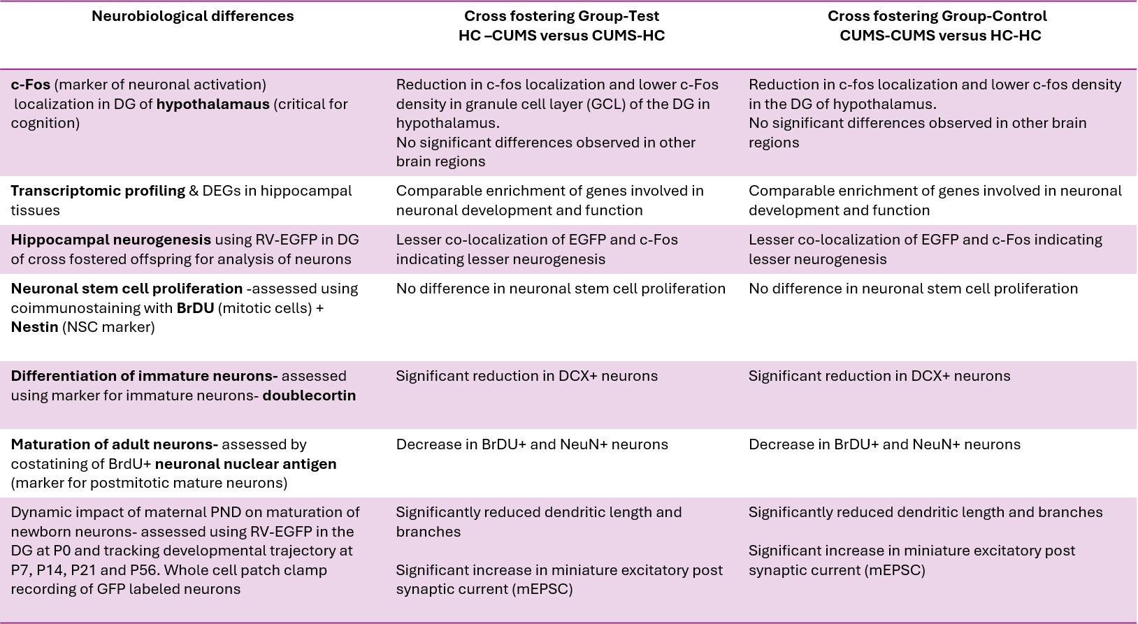
The results demonstrated that maternal PND adversely affected the maturation and synaptic integration of newborn neurons, with these alterations persisting into adulthood. This suggests that exposure to an adverse postnatal maternal environment can have long-lasting consequences for neuronal development and brain function.
What did the researchers find in breastmilk?
The study compared breastmilk samples from 91 women using small RNA sequencing: 40 diagnosed with PND and 51 healthy control mothers. The researchers identified differences in the miRNA profiles of the two groups. Among the miRNAs that were increased in the PND group, three stood out: miR-15a-5p, miR-15b-5p and miR-205-5p. The same three miRNAs were elevated in breastmilk from stressed mice used as a model of PND.
This human sample analysis shows an association between PND and changes in breastmilk miRNA content. On its own, however, it cannot establish whether these changes affect the development of a child’s brain. To investigate a possible cause-and-effect relationship, the researchers carried out a series of experiments in mice.
Could breastmilk sEVs reach the developing brain?
The researchers used labelled sEVs to follow their movement in nursing offspring. They detected signals from mammary-gland-derived sEVs in several brain regions, including the hypothalamus and hippocampus. In the hippocampus, some of the signal was found in cells expressing NeuN, a marker commonly used to identify neurons.
They also found higher levels of the mature forms of the three PND-associated miRNAs in the brains of offspring exposed to PND-associated breastmilk. The primary transcripts of these miRNAs were not similarly increased. This pattern supports the possibility that mature miRNAs had been delivered from outside the offspring’s brain, rather than being produced in greater amounts by the brain itself.
What happened when mouse pups received sEVs from human breastmilk?
One of the study’s key experiments tested whether sEVs from human breastmilk could influence mouse development. Healthy mouse pups were given human breastmilk sEVs from either mothers with PND or healthy control mothers throughout the nursing period. The pups were raised by healthy foster mothers, helping the researchers reduce the influence of differences in maternal care.
Pups that received PND-derived sEVs performed less well in tests of learning and memory than pups given sEVs from healthy control mothers. They also showed increased levels of the three miRNAs in the brain and changes in the development of neurons. By contrast, pups given breastmilk fractions from which sEVs had been removed did not show the same behavioural pattern.
These results suggest that the sEV-containing fraction of breastmilk can influence outcomes in this mouse model. They do not show that the same effects occur in human infants.
When these microRNAs were overexpressed in mammmary cells of healthy foster dams, the offspring fostered by them displayed characteristics of pups fostered by CUMS dams especially decrease in dendritic length and complexity. On the other hand, since CUMS dam already expressed these microRNAs at elevated levels, the researchers used microRNA sponges to reduce the expression of these RNAs specifically. When CUMS dams fostered healthy pups this time round, the cognitive and emotional deficits displayed previously were rescued.
How do breastmilk microRNAs travel to the offspring brain?
The researchers observed that the level of these microRNAs was elevated in the hippocampus of the offspring brain, however the level of primary transcript was the same. This indicates that the mRNAs were transferred exogeneously to the brain.
To test whether small extracellular vesicles of the mammary glands are involved in transporting miRNAs as cargo to the offspring brain, researchers used CD63-GFP model to label sEVs in mammary gland. CD63 is a marker of extracellular membranes and GFP (Green flourescent protein) is used for tracking. After 3 days of lactation, they observed strong GFP signal in the offspring hippocampus, cortex and hypothalamus. They observed colocaliztion with neuronal marker NeuN in the hippocampus.
To further confirm the role of sEVs as independent causative factor linking maternal PND to offspring cognitive effects, the researchers purified sEVs from human breastmilk. Since characteristics of miRNAs are conserved between humans and rodents, the breastmilk purifed sEVs from humans were administered to mouse pups which were then fostered by healthy dams. PND-sEV treated pups showed the same cognitive and emotional deficits as obserevd previously with elevated levels of all three miRNAs in the hippocampus whereas the human breastmilk component devoid of the sEVs did not elicit the same response.
How might these miRNAs affect brain development?
The researchers then investigated how the three miRNAs might influence the brain. Their findings pointed to a receptor called metabotropic glutamate receptor 8 (mGluR8). This receptor helps regulate communication between neurons and was identified as a direct target of the three miRNAs.
The proposed pathway involves the supramammillary nucleus (SuM), a region in the hypothalamus that sends signals to the hippocampus. The researchers found that the PND-associated miRNAs reduced mGluR8 levels in neurons that connect the SuM to the hippocampal dentate gyrus (DG). The DG is involved in learning and memory and is one of the brain regions where new neurons are generated.
When mGluR8 levels were reduced, the researchers observed abnormal neuronal activity in the DG. Calcium events occurred more frequently, although their individual amplitude did not increase. The authors describe this as a 'high-frequency, low-amplitude' activity pattern. They suggest that this altered signalling may interfere with the way newborn neurons develop and become integrated into existing brain circuits.
What does this mean for mothers and babies?
The study highlights a potential communication pathway between mother and infant. It suggests that maternal postnatal mental health may be associated not only with changes in caregiving and the home environment, but also with changes in the biological contents of breastmilk.
However, the findings should be interpreted carefully. The human part of the study identified differences in breastmilk miRNAs, while most of the experiments testing how those miRNAs affect the brain were carried out in mice. Giving human breastmilk sEVs to mouse pups is a useful way to investigate biological effects, but it cannot tell us whether the same outcomes occur in human infants or how large any such effects might be.
There are also limitations within the animal work. Although the researchers assessed behaviour in both male and female offspring, most of the detailed brain and molecular experiments were performed in males. Further studies will be needed to understand whether the proposed pathway operates in the same way in females and to establish how relevant it is to human development.
Importantly, this research is not evidence that mothers with PND should stop breastfeeding. Breastfeeding has established nutritional and other benefits, and this study does not assess the overall balance of benefits and risks for human infants. Nor does it suggest that mothers are responsible for the effects of depression. Rather, it reinforces the importance of recognising and treating PND and of supporting maternal wellbeing.
The bigger picture
This research opens an interesting new area of investigation into how maternal mental health may be linked to infant brain development. By identifying specific miRNAs in breastmilk sEVs and tracing a possible pathway involving mGluR8 and the hippocampus, the researchers provide a mechanistic explanation that can now be tested further.
The next step is to determine whether these findings translate to human infants, whether the identified miRNAs can reliably indicate changes associated with PND, and whether supporting maternal mental health can influence these biological pathways. For now, the central message is that caring for maternal mental health is important — for mothers themselves and for the wider family environment in which infants develop.
Reference
Zexu Wei, Ranran Yu, Junnan Xu, Chenyu Tao, Aixia Zhang, Longjie Li, Xiaoshan Zhang, Jin He, Jiajun Yu, Xuhuang Xiao, Jiahui Guo, Tong Wang, Wei Gong, Yanbo Wang, Chen-Yu Zhang, Dijun Chen, Zhang-Peng Chen, Chenbo Ji, Xi Chen,
Postnatal depression-associated breastmilk extracellular vesicles mediate intergenerational effects on offspring neurodevelopment, Cell Metabolism,
2026, https://doi.org/10.1016/j.cmet.2026.09.023