The science of antidepressants just got deeper.


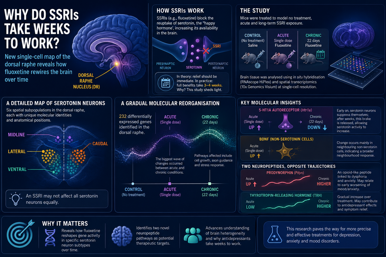
SSRIs- selective serotonin reuptake inhibitors- like fluoxetine (Prozac), sertraline, and citalopram are among the most widely prescribed medications for depression, anxiety, and mood disorders. They work by blocking the reabsorption of serotonin, often called the 'happy hormone', back into nerve cells, temporarily raising serotonin levels in the brain. In theory, this should relieve symptoms of depression almost immediately. In practice, however, most patients need to take SSRIs consistently for 3–4 weeks before experiencing their full therapeutic effect. This is a paradox that remains a central question in antidepressant research.
Serotonin is produced by specialised brain cells called serotonergic neurons, clustered in a small but powerful region of the brain known as the dorsal raphe nucleus (DR). Antidepressants are known to trigger changes in gene activity across the brain, with particularly pronounced effects in this region. Yet despite decades of research, scientists still don't fully understand how these drugs affect the distinct types and subtypes of serotonergic neurons in the brain.
New research from Stockholm University addresses this gap using state-of-the-art methods to map gene expression changes at single-cell resolution with high precision.
The study compared three groups of mice: one receiving saline solution as a control, one given a single dose of fluoxetine, and one treated with fluoxetine daily for 22 days, designed to mirror no treatment, acute and long-term treatment, respectively. The brain tissue was harvested and cryosections prepared for in situ hybridisation and spatial transcriptomics.
The study identified six distinct spatial subpopulations of serotonin neurons in the dorsal raphe, each with unique molecular identities and anatomical positions- midline, caudal, lateral, and ventral zones. This matters because it means an SSRI may not affect all serotonin neurons equally.
232 differentially expressed genes were identified in the dorsal raphe. The biggest wave of changes occurred between the acute and chronic conditions — suggesting the brain undergoes a major molecular reorganisation over the course of treatment, affecting signalling pathways linked to cell growth, nerve fibre guidance, and stress response.
The 5-HT1A autoreceptor gene (Htr1a) was upregulated after a single dose of fluoxetine; however, chronic treatment decreased its expression. This supports a long-standing theory: early on, serotonin neurons suppress themselves in response to the drug; only after weeks does this brake release, allowing serotonin activity to increase properly. This may help explain why antidepressants take so long to work.
BDNF (brain-derived neurotrophic factor), linked to neuroplasticity and mood regulation, was among the top upregulated genes after acute treatment. Interestingly, this change occurred mainly in nearby non-serotonin cells, suggesting SSRIs trigger a broader neighbourhood response beyond the cells they directly target.
Interestingly, the researchers identified significant changes in two neuropeptides, whose expression changed in opposite directions over the course of treatment. Prodynorphin (Pdyn) is an opioid like peptide associated with receptors linked to dysphoria and anxiety. The worsening of anxiety and mood that some patients experience when they start fluoxetine treatment may be associated with the increase in the level of this neuropeptide.
The second neuropeptide identified, thyrotropin-releasing hormone (TRH), is recognised for its involvement in emotional regulation. TRH levels increased gradually over the course of fluoxetine treatment, indicating a potential role in mediating the antidepressant effects of the drug and the subsequent reduction of depressive symptoms.
By identifying two previously unrecognised neuropeptides that are modulated by fluoxetine, this study provides a foundation for future investigations into novel therapeutic targets, potentially enabling the development of more precise and effective pharmacological interventions.
Furthermore, it advances our understanding of the brain's cellular and molecular heterogeneity, highlighting how different cell populations and genetic pathways may respond to antidepressant exposure.
Snapshot
Reference
Henningson, C., Mlost, J. & Pollak Dorocic, I. Effects of SSRIs on the spatial transcriptome of dorsal raphe serotonin neurons. Mol Psychiatry (2026). https://doi.org/10.1038/s41380-026-03644-x